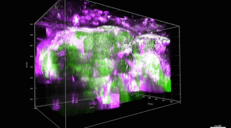
Three-photon microscopy, a technique for in-vivo imaging pioneered at Cornell Engineering, has been increasingly adopted for probing neural activities beyond the typical two-photon imaging depth. In the journal Optica, Professor Chris Xu and Postdoctoral Associate Tianyu Wang of the School of Applied and Engineering Physics outline the unique properties that differentiate three-photon microscopy from two-photon microscopy for in vivo imaging in biological samples, especially in the mouse brain.
 They present a systematic summary of the optimization of three-photon imaging parameters for neural imaging, based on their effects on calcium imaging quality and perturbation to brain tissues. Furthermore, they review the existing techniques for volumetric imaging and discuss their prospects in mesoscale three-photon imaging in deep tissue.
The review paper, "Three-photon neuronal imaging in deep mouse brain," published online Thursday, Aug. 6, 2020.